
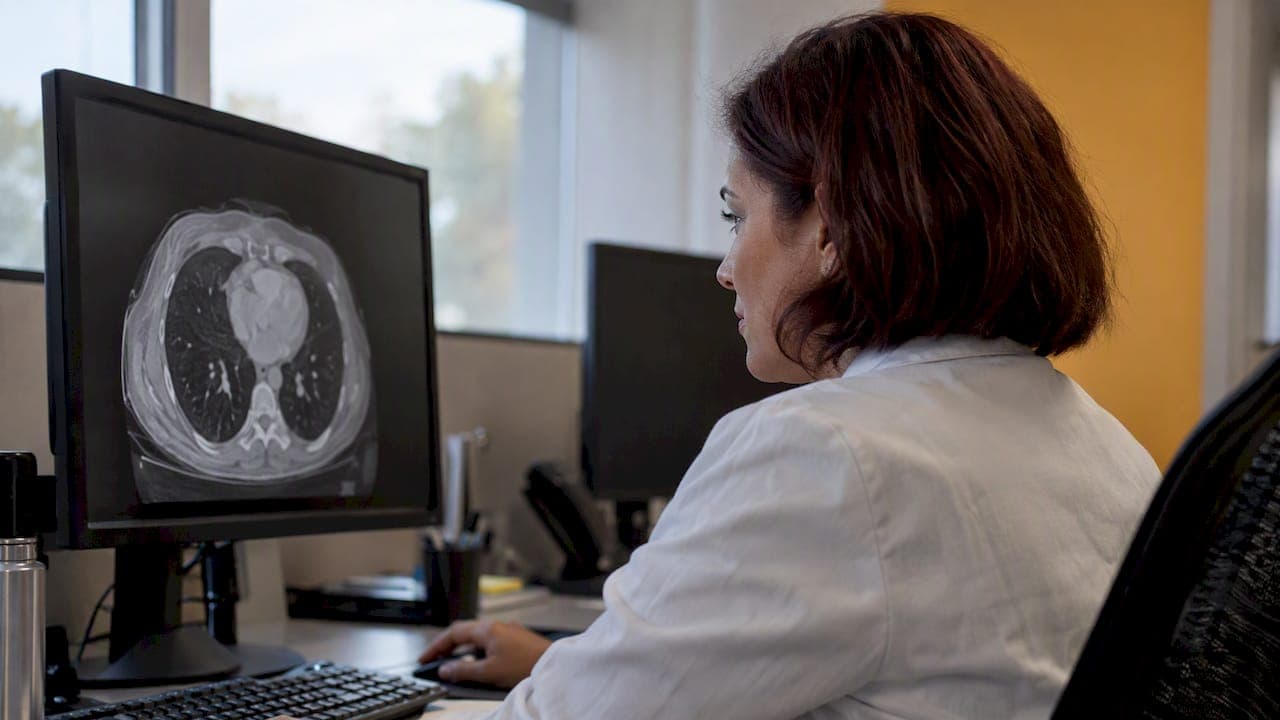
There is no single "convert" button for DICOM to SDTM. DICOM is the standard for acquiring, storing and describing medical images. SDTM is the model for tabulating clinical trial data. Moving from one to the other means deciding which imaging facts matter to your protocol, translating them into the right SDTM domains, and keeping a verifiable link back to the source image. Done well, the result is a dataset a reviewer can trust and trace. Done late, it becomes a scramble at submission.
This guide is for CRO data managers, sponsor clinical operations teams and imaging leads who have to make that mapping work. It covers what the conversion really is, how oncology imaging lands in TU, TR and RS, how to design traceability, and what to scope before anyone writes code.
TL;DR
- DICOM to SDTM is a mapping and standardization workflow, not a field-for-field export of DICOM tags. You decide which trial-relevant findings belong in SDTM and keep a link to the source.
- In oncology, lesion identification goes to TU, lesion measurements to TR, and overall response to RS. Imaging procedure details can go to PR when the procedure itself matters.
- FDA recommends implementing SDTM before the study begins, with a complete define.xml and a reviewer's guide, so plan the mapping at protocol stage.
- DICOM Structured Reports are far easier to transform than free-text radiology reports.
- Cost and timeline cannot be quoted generically. Scope them from a sample dataset and a draft mapping specification.
What "DICOM to SDTM" actually means
DICOM objects carry pixel data plus a large set of metadata: patient and study identifiers, modality, acquisition parameters, series and instance identifiers. Some objects, such as Structured Reports, also carry measurements and findings. SDTM, by contrast, is organized into domains of observations about subjects in a study.
The SDTM Implementation Guide (SDTMIG) guides the organization, structure and format of standard clinical trial tabulation datasets. It is not a DICOM conversion specification. Nothing in it tells you how to parse a DICOM file. That part is your design.
CDISC offers a helpful way to think about it in its article on why an image is like a specimen. A lab sample is a surrogate for the subject, and so is an image. The useful SDTM record is the standardized observation, linked to when and how the image was produced. A wholesale dump of scanner metadata is not the goal.
In practice, a production workflow does five things:
- Extracts trial-relevant metadata and findings from DICOM objects or Structured Reports.
- Normalizes terminology and units.
- Maps results into the applicable SDTM domains.
- Validates conformance and relationships between records.
- Delivers datasets with metadata and traceability documentation.
Decide what is trial-relevant first
The most common mistake is starting from the files instead of the protocol. Ask these questions before you open a single DICOM header:
- Which imaging assessments support the primary and secondary endpoints?
- Who interprets the images: the site investigator, a central reader, or both?
- Is the finding quantitative (a diameter in millimeters), qualitative (present or absent), or a derived response category?
- Does the procedure itself matter to the analysis, for example a scan date that anchors a visit window?
The answers determine which domains you need. A study with a single imaging-based response endpoint needs a very different design from one that collects dozens of structural measurements.
FDA frames this as a process question as well as a data question. Its imaging endpoint guidance recommends standardized acquisition, display, archiving and interpretation processes when imaging supports a primary endpoint or part of one, because reducing variability protects data quality. The guidance names DICOM and PACS as established clinical imaging standards, and then asks for trial-specific process standards on top of them.
The practical lesson is reassuring and sobering at once. A technically valid SDTM dataset cannot repair inconsistent imaging procedures or undocumented changes in interpretation. The mapping is only as strong as the imaging process behind it. Our post on turning FDA imaging guidance into site checks with an imaging charter walks through how to set that foundation at the sites.
Mapping oncology imaging to TU, TR and RS
Oncology is where most DICOM-to-SDTM work happens, so it is worth being specific. CDISC's medical imaging data standards presentation identifies three key domains:
| Domain | What it holds | Typical imaging source |
|---|---|---|
| TU (Tumor/Lesion Identification) | Each tumor or lesion, with a stable identifier and location | Lesion annotations or reader selections |
| TR (Tumor/Lesion Results) | Quantitative or qualitative results for each lesion, such as measurements | Measurements in a Structured Report or reader tool |
| RS (Disease Response) | Overall response assessments at each time point | Reader or investigator response evaluation |
The same material notes that the imaging procedure does not always need to be represented separately. When the procedure itself is relevant to the study, PR can carry it. The right choice depends on what your study actually collects, so resist forcing every imaging fact into one domain.
Keep lesion identity stable
A frequent hidden failure is separating lesion identity from lesion measurements and response. If a lesion is labeled one way at baseline and another way at week 12, TR rows cannot be tied back to TU, and the response in RS loses its foundation. Stable linking identifiers matter more than simply extracting measurements. Build the key logic first and test it across every visit before you worry about formatting.
Normalize terms and units
Readers and tools express the same finding in different words and units. Decide on controlled terminology and unit conventions in the mapping specification, and apply them in code rather than by hand. This is the same discipline that makes lab data submission-ready, and our guide to mapping CDASH to SDTM lab data with LBLOINC shows how it plays out on the laboratory side.
Structured reports make the job tractable
If your imaging vendor or reading platform can produce DICOM Structured Reports, use them. These objects encode measurements, annotations and analytic results in a structured form, so a program can read a lesion diameter directly instead of interpreting prose. Free-text radiology reports are much harder to transform and carry more risk of error and inconsistent interpretation.
DICOM also defines templates for encoding imaging reports using HL7 CDA, and supports explicit evidence references that point from a finding to the images supporting it. Those references are valuable for traceability, which is the next topic.
If you are choosing a reporting format for a new study, we cover five practical checks in DICOM SR for trials and traceable SDTM mapping.
Design traceability from the start
A reviewer, auditor or internal QA lead should be able to pick any SDTM record and answer four questions: which image or structured-report object did this come from, which imaging event was it, who evaluated it, and which transformation rule produced the value.
That chain is the difference between a dataset and an evidence trail. The source standards support it: DICOM provides identifiers and evidence references, and CDISC notes that image identifiers can be represented through SDTM reference-ID variables. The final row should never be an isolated number.
A workable design usually includes:
- Source references. Study, series and instance identifiers, or a Structured Report object identifier, stored alongside each derived record or in a controlled linking table.
- Event context. Visit, date and procedure, so each finding sits in the right place on the subject's timeline.
- Evaluator identity. Whether the value came from a site reader, a central reader or an algorithm, and which reader if several.
- Rule versioning. The version of the mapping specification and code that produced each record, so changes are never silent.
The broader principle is the same one any regulated team applies to its own records: a log is only useful if it proves something. For a plain-language look at what auditable trails in healthcare should demonstrate, that discussion carries over well to imaging data lineage.
Plan before the study starts
The conversion should be designed before study conduct, not improvised at submission. For a U.S. submission, FDA's Study Data Technical Conformance Guide recommends implementing SDTM before the study begins, following the applicable SDTMIG and the FDA Data Standards Catalog. It also expects a complete define.xml and an appropriate reviewer's guide with the datasets. FDA applies business and validator rules to assess conformance and reviewability, so your validation should anticipate them.
Lock your versions early
CDISC stresses that each SDTMIG is tied to a particular SDTM model release, and that models evolve cumulatively. Choose the SDTM, SDTMIG, controlled terminology and FDA-supported versions at the outset. Otherwise a mapping that looks correct can drift out of line with the version you declare in the submission.
When the guide does not answer your question
Imaging studies sometimes produce data the implementation guide does not clearly address. FDA's guide recognizes this. The sound approach is to document your rationale, record the open decision in the study data reviewer's guide, and, for application-specific uncertainty, raise it with the review division. Inventing a mapping quietly and hoping it passes is the one path to avoid.
A practical workflow
Here is a sequence that teams can adapt to their own protocol:
- Read the protocol and imaging charter. List every imaging-derived endpoint and assessment.
- Choose the source format. Prefer Structured Reports or a reading platform export with structured fields over free text.
- Draft the mapping specification. For each variable, state the source element, the transformation, the target domain and the controlled terminology.
- Build a sample dataset. Use a handful of subjects across several visits, including awkward cases such as missing scans, re-reads and new lesions.
- Run validation. Check conformance, relationships between TU, TR and RS, and reconciliation against the reader's source record.
- Document decisions. Feed unresolved choices into the reviewer's guide and define.xml metadata.
- Move to a controlled pipeline. Version the code, repeat the validation at each data cut, and keep the traceability links.
Steps 3 and 4 are where most of the risk drops. A mapping specification tested on a small, difficult sample will tell you more than a month of planning in the abstract.
Common failure points
- Dumping DICOM tags. Scanner metadata is rarely an SDTM observation. Map findings and context, not every header.
- Losing lesion continuity. Identifiers change between time points and the link to TU breaks.
- Mixing evaluators. Site and central reads land in the same rows without a way to tell them apart.
- Skipping provenance. Values arrive without a path back to the source object, which makes queries painful and audits slow.
- Mapping at the end. Specification, terminology and version choices are made under submission pressure.
- Treating the file as the fix. Format conformance does not repair an inconsistent imaging process.
Our perspective
We see the same pattern across research programs: the teams that struggle with imaging data are rarely short of technology. They are short of agreement, early, on who owns each decision. Imaging, data management, biostatistics and the laboratory all touch the same subject timeline, and each one tends to build its own view of it.

That is why we think of imaging and laboratory results as one data problem for the sponsor. A central partner that handles specimen logistics, testing and standards-aligned data delivery can apply the same discipline to every source: stable identifiers, consistent terminology, documented rules and datasets that arrive ready for analysis. Our approach to analysis-ready lab data follows a standards-first sequence, and the same habits carry over to imaging.
Our advice is simple and hopeful. Imaging data does not have to be the last, hardest piece of a submission. Decide what the protocol needs, pick structured sources, lock versions, test on a difficult sample, and keep every value traceable. Do those five things early and the conversion becomes a routine, reviewable process.
— Kohealth Labs
Where KoHealth Labs fits
If you are building a trial data package that combines laboratory and imaging information, we can help you plan how those results reach your team in a consistent, standards-aligned form. Our clinical trials and research laboratory services cover specimen collection and management, testing, biomarker analysis and data delivery designed around study-specific requirements. Learn more on our clinical trials page, or see how our process works before you reach out with your protocol.
What to scope before asking for a quote
There is no universal price or fixed turnaround for DICOM-to-SDTM conversion, and FDA's guidance defines submission and documentation expectations rather than commercial pricing or project length. Be wary of any generic figure. These factors drive the real effort:
- Imaging modality and the number of different assessments.
- Whether findings are structured or must be extracted from text.
- Number of sites and subjects.
- Endpoint complexity, such as multi-reader adjudication.
- Terminology mapping and validation requirements.
- One-time migration versus an ongoing, controlled pipeline with repeated data cuts.
The best way to get a reliable estimate is to share a sample dataset and a draft mapping specification. Both let a provider see the real complexity, and both give you something concrete to compare.
FAQ
Can DICOM be converted directly to SDTM?
Not as a one-step export. DICOM holds imaging data and metadata, while SDTM holds standardized trial observations. You extract the trial-relevant findings, normalize them, map them to domains such as TU, TR, RS or PR, and keep links back to the source objects.
Which SDTM domains are used for imaging in oncology?
TU holds lesion identification, TR holds lesion measurements or findings, and RS holds overall disease response. PR can represent the imaging procedure when it is relevant to the study. The choice depends on what your protocol collects.
When should DICOM-to-SDTM mapping be planned?
Before the study begins. FDA recommends implementing SDTM before study start, and submitting a complete define.xml and a reviewer's guide with the datasets. Late mapping tends to produce version mismatches and undocumented decisions.
Is a DICOM Structured Report required?
No, but it helps a great deal. Structured Reports encode measurements and annotations in a machine-readable form, which is much easier to transform than free-text radiology reports. Whatever the source, keep the source object and provenance in your mapping.
How much does DICOM-to-SDTM conversion cost?
There is no standard price. Cost and timeline depend on modality, structure of the findings, site and subject counts, endpoint complexity and validation needs. Scope it from a sample dataset and a draft mapping specification.
Sources
- SDTMIG | CDISC
- Why is an Image like a Specimen? | CDISC
- Medical Imaging Data Standards, Automation & Analysis | CDISC
- Clinical Trial Imaging Endpoint Process Standards Guidance for Industry | FDA
- Study Data Technical Conformance Guide | FDA
- PS3.20 | DICOM Standard
For more information about our diagnostic testing, clinical research laboratory services, or partnership opportunities, visit https://kohealthlabs.com or contact KoHealth Labs at (469) 792-8439 and ask for Amy Patterson.