
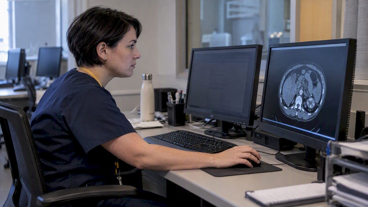
Use DICOM Clinical Trial Identification attributes together with TID 1500-based Structured Report templates to capture trial measurements consistently while preserving a clear link back to the source images. Tag each study with Protocol, Subject, Site, and Time Point identifiers, then extract and map those SR values into CDISC SDTM domains such as TU, TR, and RS so the data arrives submission-ready.
TL;DR:
- Clinical Trial Identification attributes are optional; standardize Protocol, Subject, Site, and Time Point tags, and do not mistake consent metadata for image deidentification.
- TID 1500 measurement groups need unique tracking UIDs and source SOP Instance references so auditors can trace each value to its image across visits.
- Choose one tagging point, such as scanner, PACS, site workstation, or central coordination, and retain the SOP Instance UID for audit traceability.
- For submission, extract SR values, normalize units and coding, map them to TU, TR, and RS, then run conformity and RECIST consistency checks.
- Codes that separate anatomy, findings, and modifiers usually map more cleanly into AIM and SDTM, while combined codes simplify initial reporting.
What DICOM Provides for Clinical Trials: Identification Modules and Key Tags
The DICOM standard includes an optional Clinical Trial Identification extension built specifically for research use. It lets a trial team group imaging studies under a single protocol regardless of which site or scanner produced them, which matters when a study spans multiple sites and imaging vendors.
The attributes trial teams should plan for in every SR template include:
- Clinical Trial Protocol ID (0012,0020): identifies which study protocol an image or report belongs to.
- Clinical Trial Subject ID (0012,0040): ties the instance to a specific enrolled participant.
- Clinical Trial Site ID (0012,0030): records which site acquired or submitted the data.
- Clinical Trial Time Point ID (0012,0050): marks the visit or interval, essential for longitudinal comparison.
These modules are optional in the base standard, which is exactly why Supplement 70 exists: without an explicit agreement to use them, multi-site trials end up with inconsistent metadata that makes grouping and audit trails much harder to reconstruct later. Supplement 70 also defines a Consent for Clinical Trial Use Sequence (0012,0083), which records permission to distribute an instance but does not substitute for a formal de-identification process, a distinction worth keeping straight from the start.
Using TID 1500 as the SR Measurement Report for Trial Data
TID 1500, the Measurement Report template, functions as the top-level container for quantitative imaging findings in a DICOM SR document. Inside it, measurements are organized into nested measurement groups, each carrying a unique tracking UID that lets a reviewer follow the same lesion or anatomical region across multiple time points in a longitudinal trial.
Each group can hold quantitative values like lesion diameter or volume as well as categorical findings like response classification, and every measurement should reference the source SOP Instance it was derived from. That reference is what lets an auditor trace a submitted number back to the exact image slice it came from, a requirement most trial sponsors will ask about directly.

One design choice affects everything downstream: whether findings are pre-coordinated (using a single code that already combines anatomy, finding, and modifier) or post-coordinated (building the same meaning from separate codes). Pre-coordination tends to simplify initial reporting, while post-coordination usually transforms more cleanly into other formats, including AIM and eventual SDTM mapping. Deciding this early avoids a costly remapping exercise later in the study.
Where to Add Clinical-Trial Metadata in the Imaging Workflow
Clinical Trial Identification attributes can be inserted at several points, and the right choice depends on how much control a trial exercises over site equipment. Common insertion points include:
- Scanner level: attributes are added at acquisition, before the image ever leaves the site.
- PACS ingestion: metadata is added as studies are received into the site's archive.
- Local site workstation: a technologist or research coordinator adds trial tags manually or through site software.
- Clinical Trial Coordinating Center (CTCC): attributes are added centrally after images arrive from all sites.
Adding these attributes does not inherently require changing the SOP Instance UID, which matters because it keeps the audit trail intact: the same instance can be identified as the one originally acquired, even after trial metadata is layered on at a later workflow point.
For blinded central reads, manage de-identification separately from adding Clinical Trial Identification attributes, using a controlled, documented process. Our guide on DICOM de-identification without losing research value walks through the attribute-level tradeoffs in more detail.
Pro Tip: Lock down which workflow point owns CTI tagging before the first site goes live. Retrofitting it across sites that already have inconsistent metadata is far more work than agreeing on the insertion point up front.
Mapping DICOM SR Measurements to CDISC SDTM Domains for Submission
Regulatory submissions expect data in CDISC SDTM format, not raw DICOM tags, which means every imaging-derived measurement eventually needs to land in a standard domain. The most relevant domains for imaging trials are Tumor/Lesion Identification (TU), Tumor/Lesion Results (TR), and Disease Response (RS), each built to hold a specific slice of what a radiologist records in an SR Measurement Report.
A practical pipeline looks like this: extract DICOM tags and SR measurement values, normalize units and coding systems, map the normalized values to the correct SDTM variables, run conformity and RECIST consistency checks, then generate the submission datasets. Each step depends on the one before it, so an error introduced at extraction propagates all the way through to the final dataset if it isn't caught early.

Automation helps here more than almost anywhere else in the pipeline. Tools modeled on CTIMS-style pre-screening and DICOM-to-SDTM conversion utilities reduce the manual transcription errors that come from copying tumor measurements by hand between systems. Our breakdown of structured diagnostic data regulatory submission requirements covers the documentation expectations that sit alongside this mapping work.
Practical Checklist: Configure, Test, and Distribute a Trial-Specific SR Template
Getting a template from design to multi-site use involves several stages:
- Define the template. Set the root container title, required child content items, constrained value representations, required Clinical Trial Identification attributes, and tracking UID rules, modeled on the TID 1500 structure.
- Test locally. Generate SR instances from sample images, confirm every measurement correctly references its source SOP Instance, and validate that measurement-group UIDs behave as expected across repeat scans.
- Version and distribute. Export the finished template profile, assign it a version, and send it to each site with install instructions and a QA checklist.
- Validate. Run automated tag-extraction tests and SDTM mapping checks, then perform sample blinded reads to confirm traceability and de-identification handling work as designed.
- Document consent and privacy handling. Include the Consent for Clinical Trial Use Sequence in the SR where required, but manage de-identification separately through confidentiality profiles rather than folding the two processes together.
Our radiology data QC guidance covers automated tag-extraction testing in more depth, useful once a template moves from pilot to full site rollout.
Integrating Imaging-Ready SR Templates Into Trial Operations
We work alongside research sponsors and CROs to bundle laboratory and imaging-ready data for active studies, which includes LIS and EHR integration so trial measurements reach a sponsor's systems without a manual handoff. That integration work surfaces the same traceability questions template design raises: where metadata gets added, how it's versioned, and how it's validated before a site ever submits data.
For teams standing up a new SR template, we offer onboarding support. If your trial team is weighing template design choices, we’re a reasonable place to start that conversation.
— Kohealth Labs
How KoHealth Labs Supports Clinical-Trial Imaging Data
Configuring a compliant SR template is only part of the operational picture. We support research sponsors and CROs with:
- Specimen logistics and courier pickup.
- LIS and EHR integration.
- Regulatory-aligned data delivery built around the traceability standards trial sponsors expect.
If your team needs a lab and imaging-data partner, visit our clinical trials services page to learn more.
FAQ
What is DICOM SR and why does it matter for clinical trials?
DICOM SR, or Structured Report, is a standardized format for recording imaging findings like measurements and classifications in a machine-readable way, rather than as free text. For trials, it matters because templates like TID 1500 keep measurement data structured and traceable across sites, visits, and readers.
Are DICOM Clinical Trial Identification attributes mandatory?
These attributes are optional in the base DICOM standard, but they can provide a way to group studies by protocol, subject, site, and time point, as described in Supplement 70. Without them, grouping and auditing imaging data across sites becomes considerably harder.
How do DICOM SR measurements get into an SDTM submission dataset?
Measurement values are extracted from the SR, normalized for units and coding, then mapped into SDTM domains such as TU, TR, and RS using CDISC's implementation guidance. Automated extraction and mapping tools reduce the manual transcription errors common in this process.
Does adding trial metadata require de-identifying images first?
No, adding Clinical Trial Identification attributes and de-identifying an image are separate processes governed by different parts of the standard. Recommended practice for blinded reads is to de-identify first, then apply trial metadata to the de-identified instance under a controlled workflow, as outlined in the Clinical Trial Study Module.
Where should trial metadata be added in the imaging workflow?
Metadata can be added at the scanner, during PACS ingestion, at a site workstation, or centrally at a Clinical Trial Coordinating Center, and the choice depends on how much control the trial has over site equipment. Adding it at any of these points does not require changing the SOP Instance UID, which keeps the audit trail intact.
Sources
For more information about our diagnostic testing, clinical research laboratory services, or partnership opportunities, visit https://kohealthlabs.com or contact KoHealth Labs at (469) 792-8439 and ask for Amy Patterson.