
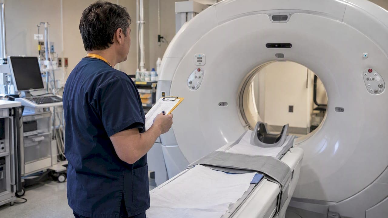
An imaging charter is the document that spells out how images are acquired, transferred, interpreted and archived in a trial, so it needs to be drafted as soon as imaging touches a key endpoint. Drafting the charter early gives the team time to define imaging processes before the trial begins. Your first move: convene your imaging lead, a medical physicist and your data manager to start the draft together.
TL;DR:
- When imaging supports eligibility or a primary endpoint, define trial specific process standards; FDA recommends a separate charter when those standards are extensive.
- Specify modality parameters, acceptable ranges, equipment and software versions, DICOM transfer, deidentification, naming, resubmission rules, and imaging variables sent for analysis.
- Qualify sites through equipment attestations and machine certification, then use phantom images, calibration, periodic performance checks, automated flags, and visual review to control variation.
- Validate required metadata at upload, automate and log deidentification, and set submission windows with reminders to prevent incomplete records and late image transfers.
- Choose central or site reads according to trial design, limit visible clinical data, qualify readers through test reads, and document adjudication decisions with reasons.
When do you need an imaging charter?
Routine diagnostic imaging and trial imaging can have different process needs. A charter can document trial-specific processes for acquiring, interpreting and archiving images.
Imaging typically supports several stages of a study:
- Screening and baseline assessment
- Quantitative response measurement
- Safety monitoring
- Imaging biomarkers, where precision across the whole imaging chain matters, as a review of quantitative imaging biomarkers explains
Consider documenting imaging processes when imaging supports eligibility or an endpoint. The FDA guidance focuses on process standards when imaging assesses a primary endpoint or one of its components. For a deeper look at scoping imaging early, see our trial designer's guide to integrated radiology.
What belongs in an imaging charter?
Think of the charter as a companion to the protocol, written with the same rigor. Adapt this checklist to your study:
- Executive summary: the role of imaging, the endpoints it supports, and the imaging variables delivered to analysis.
- Acquisition specifications: per modality, the sequences, slice thickness, contrast timing, reconstruction parameters and acceptable ranges.
- Site requirements: equipment models, software versions, technologist attestations and phantom certification.
- Data handling: DICOM export standards, de-identification rules, study ID naming, transfer method and re-submission rules.
- Interpretation: the central reader plan, blinding procedures, reader training and adjudication.
- Quality control: phantom schedules, periodic calibration, a visual review committee and deviation logging.
- Change control: versioning, where the charter lives and how updates are approved.
Pro Tip: Define acceptable parameter ranges, not single target values. Sites can then comply without guessing, and your deviation log stays meaningful.
What does the FDA expect, and when?
The FDA's guidance on clinical trial imaging endpoint process standards focuses on acquisition, display, archiving and interpretation when imaging assesses a primary endpoint or one of its components. When those process standards are extensive, the guidance itself recommends placing them in a separate imaging charter that also describes how bias and variability are controlled.
When imaging assesses a primary endpoint or one of its components, define trial-specific imaging process standards. If those standards are extensive, the FDA guidance recommends placing them in a separate imaging charter that describes acquisition, interpretation, archiving and how bias and variability are controlled.
How do you qualify sites and keep equipment consistent?
Variability across centers is the main threat to imaging data. A multicenter quality control program describes standardized acquisition protocols, machine certification, phantom images and periodic performance evaluation as elements of a quality control program for consistent image quality.
A practical sequence:
- Send a site questionnaire and require machine attestation with vendor and software versions.
- Engage a medical physicist to define calibration, phantom protocol and acceptance criteria.
- Include machine certification, phantom images and periodic performance evaluation in the quality control program.
- Set automated flags for out-of-range parameters, backed by a manual visual review committee.
Pro Tip: Consider using phantom images as part of the quality control program to assess image quality before relying on trial images.
Specialty practices show how detailed modality-specific requirements can get, as in this clinical guide to imaging in podiatry. Our guidance on radiology data QC and audit scripts covers the monitoring side.
How should imaging data move from site to analysis?
Centralized imaging workflows can include image submission, storage and query resolution. A cancer center's practical report describes centralized workflows as a way to reduce fragmentation and improve timeliness and accuracy of imaging data management. A cancer center's practical report describes site qualification, central storage, de-identification and query resolution improving timeliness and accuracy for sponsors.

Define in the charter how images are transferred, de-identified, labeled and linked to study data. Our overview of DICOM SR for traceable SDTM mapping shows how structured reporting supports that link.
Common failure points and fixes:
- Missing metadata: validate required fields at upload and reject incomplete submissions.
- Manual de-identification: automate it and log every change.
- Late transfers: set submission windows and automated reminders.
How are images read, blinded and adjudicated?
Whether to use central or site reads depends on the trial's design and needs. Document the interpretation process and how variability is controlled in the charter.
Protect blinding by redacting clinical data or limiting the metadata readers can see. Qualify readers through training, test reads and ongoing performance monitoring, and record the results. When readers disagree, route the case to an adjudicator and log the decision, with its rationale, in the charter's documentation. Standards for quantitative measures are covered in our piece on IBSI standards for imaging biomarkers.
Who owns what, and when?
Assign owners for the imaging workflow, such as sponsor and site contacts and relevant imaging or medical physics experts. Document responsibilities and key milestones in the charter.
Typical deviations include a wrong sequence, a missing series or a late transfer. Each needs a named mitigation, such as a rescan window or a technologist retraining step. Keep a versioned charter, a change log and an audit trail so an inspector can follow every decision.
A lab partner's view: friction is the real cost
Delays in imaging trials rarely come from the science. They can come from handoffs, such as a missed pickup, an agreed file format that is missing, or queries passed between teams. When specimen and imaging logistics are coordinated, those queries shrink. Before you commit to a partner, ask how they integrate with your LIS and eCRF, how pickups work and what format deliverables arrive in. Then scope it against your protocol.
— Kohealth Labs
How we support imaging-enabled trials
We offer integrated diagnostics, specimen pickup and courier services, LIS integration and regulatory-aligned data delivery for research teams. If your study pairs imaging with laboratory endpoints, our integrated clinical diagnostics for CROs and pharma can fit around your charter. Reach out through that page or book a discovery call, and we will map your imaging and lab workflows together.
This article is general information, not a substitute for advice from a qualified doctor. Consult a qualified healthcare professional about your own circumstances before acting on anything here.
FAQ
What field of study is medical imaging?
Medical imaging sits within radiology and biomedical engineering, drawing on medical physics for image acquisition and quality. In trials, it also involves data management and biostatistics, which is why charters involve several disciplines.
What is an audit trail in clinical research?
An audit trail is a time-stamped record of who created, changed or reviewed data and why. For imaging, it documents transfers, de-identification, reads and adjudication, so regulators can reconstruct every step.
What are the four types of scans?
There is no single official list. Commonly cited modalities include X-ray, CT, MRI and ultrasound, and trials often add nuclear imaging such as PET. Your charter should define parameters for each modality your protocol uses.
When should sponsors start an imaging charter?
Start planning imaging processes when imaging is identified as central to eligibility or a primary endpoint. The FDA guidance focuses on process standards when imaging assesses a primary endpoint or one of its components.
Sources
- Clinical Trial Imaging Endpoint Process Standards Guidance for Industry | FDA
- A Review on the Use of Imaging Biomarkers in Oncology Clinical Trials: Quality Assurance Strategies for Technical Validation - PMC
- Description and implementation of a quality control program in an imaging-based clinical trial - PubMed
For more information about our diagnostic testing, clinical research laboratory services, or partnership opportunities, visit https://kohealthlabs.com or contact KoHealth Labs at (469) 792-8439 and ask for Amy Patterson.