

Integrated radiology in clinical trial design is defined as the deliberate, protocol-level alignment of imaging endpoints, acquisition standards, and analysis workflows with a study's primary clinical objectives. The role of integrated radiology in study design goes far beyond ordering scans at scheduled visits. When imaging is treated as a core evidence source from day one, rather than a secondary data stream, it produces decision-ready, reproducible results that satisfy FDA imaging biomarker qualification standards and ACR guidelines. Trials that treat imaging as an afterthought generate "less than ideal" data that blocks regulatory success. The industry term for this practice is "imaging integration," and it covers everything from DICOM standardization to central imaging review to AI-enabled workflow automation.
What is the role of integrated radiology in study design?
Imaging integration is the single most preventable source of trial delay when it is handled reactively. Early imaging planning is essential for regulatory-grade data, according to the PROVIDENT study consensus. That means defining modality, acquisition parameters, reader qualification criteria, and endpoint measurement rules before the first site is activated.
The consequences of skipping this step are measurable. Inter-reader discordance rates in oncologic trials historically range between 20–40%. A discordance rate that high means a significant portion of imaging reads cannot be reconciled, which forces protocol amendments, delays database lock, and can invalidate primary endpoints.

Tumor metrics imaging core labs address this problem directly. Centralized review enhances quality and reduces variability across multi-site trials by replacing inconsistent local reads with a single, standardized analysis process. For trial designers, this is not optional in oncology studies. It is the structural backbone of a defensible imaging dataset.
The benefits of integrated imaging extend to regulatory submissions as well. When imaging endpoints are defined early and executed consistently, the data package you submit reflects a coherent, pre-specified analysis plan. Regulators can trace every measurement back to a documented protocol decision.
Pro Tip: Define your imaging endpoints, reader qualification criteria, and DICOM transfer specifications in the protocol synopsis, not the final protocol. Changes made after site activation are expensive and often require IRB amendments.
Key steps to lock in during initial study design include:
- Select imaging modality and field strength based on the target tissue and endpoint sensitivity
- Specify acquisition parameters per scanner manufacturer to reduce site-to-site variability
- Define measurement criteria (RECIST 1.1, iRECIST, or study-specific rules) before enrollment opens
- Identify whether local reads, blinded independent central review, or both are required
- Establish data transfer, de-identification, and metadata tagging requirements upfront
How do AI-enabled platforms change imaging workflows?
Modern AI-driven radiology platforms reduce reporting turnaround times to approximately 30 minutes. That speed matters in trials where imaging findings trigger dose modifications or treatment discontinuation decisions. Waiting days for a read is not a workflow inconvenience. It is a patient safety issue.
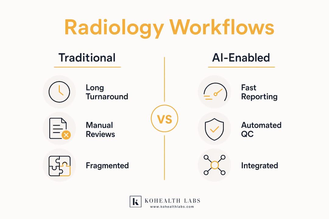
These platforms handle image ingestion, automated triage, structured reporting, and quality control within a single integrated environment. Complete radiology workflow automation, classified as Level 5 on the Radiology AI Maturity Map, means that AI flags acquisition errors, routes studies to qualified readers, and generates structured reports without manual handoffs. That removes the variability introduced by site-level processes.
The table below compares traditional and AI-enabled radiology workflows across the dimensions that matter most to trial designers.
| Workflow dimension | Traditional radiology | AI-enabled integrated radiology |
|---|---|---|
| Turnaround time | 24–72 hours per read | Approximately 30 minutes |
| Quality control | Manual, post-read | Automated, at acquisition |
| Structured reporting | Inconsistent across sites | Standardized templates |
| DICOM compliance | Site-dependent | Protocol-enforced |
| Scalability | Limited by reader availability | Scales with study volume |
DICOM standardization sits at the center of this shift. Imaging is evolving from a secondary diagnostic tool to a primary evidence driver, enabled by AI and workflow integration. DICOM metadata carries acquisition parameters, scanner identifiers, and sequence details that are essential for post-processing reproducibility. Without enforced DICOM compliance, two sites running identical protocols can produce non-comparable datasets.
Why does interdisciplinary collaboration determine imaging success?
Successful imaging integration requires aligning modality, endpoints, and analytical methods with developmental goals from the outset. That alignment does not happen when imaging experts are brought in after the protocol is drafted. It requires a cross-functional team working together from the study conception phase.
The PICO framework (Population, Intervention, Comparator, Outcome) gives interdisciplinary teams a shared structure for aligning imaging markers with clinical priorities. When the imaging scientist maps the "Outcome" component of PICO to a specific measurable imaging endpoint, the clinical team and data scientists can build their analysis plan around a concrete, pre-specified target.
A practical structure for your imaging integration team looks like this:
- Principal investigator defines the clinical question and primary endpoint in plain language before any imaging modality is selected.
- Imaging scientist or radiologist translates the clinical question into a measurable imaging endpoint and specifies the acquisition protocol.
- Data scientist or biostatistician confirms the endpoint is quantifiable, reproducible, and powered appropriately in the sample size calculation.
- Regulatory affairs lead reviews the imaging endpoint against FDA imaging biomarker qualification guidance and relevant precedent studies.
- Clinical operations lead maps the imaging schedule to site capabilities and confirms DICOM transfer infrastructure before site qualification.
This sequence prevents the most common failure mode: a clinically meaningful endpoint that cannot be measured reliably with the available imaging infrastructure. Bringing the data scientist into the conversation after the protocol is finalized routinely produces endpoints that are clinically sound but statistically underpowered.
Pro Tip: Schedule a dedicated imaging design meeting within the first two weeks of protocol development. Include your imaging core lab contact at that meeting. Decisions made in that session will shape every imaging-related protocol amendment you avoid later.
What are the common challenges in implementing integrated radiology?
The most damaging challenge is inter-reader variability. Unblinded local reads in lung cancer trials produce an average 32.9% inter-reader discordance, based on an analysis of 1,833 patients across six trials by 71 radiologists. That figure means roughly one in three reads would be classified differently by a different reader. Blinded independent central review is the direct solution, and it should be specified in the protocol, not added as a corrective measure after enrollment.
Data interoperability is the second major challenge. Failures in image flow or de-identification can invalidate entire data batches and delay trials. Pre-activation testing of the full imaging pipeline, from acquisition at the scanner to receipt at the central lab, catches these failures before they affect real patient data.
Common implementation challenges and their solutions include:
- Scanner protocol variability across sites: Require site qualification imaging with phantom scans before enrollment. Reject sites that cannot meet acquisition specifications.
- De-identification errors: Use automated de-identification software with audit logs. Manual de-identification introduces errors that expose patient data and corrupt metadata.
- Metadata loss during transfer: Enforce DICOM tag verification at the receiving end. Missing tags make images unreadable by analysis software.
- Reader drift over long trials: Schedule periodic reader calibration sessions using reference cases. Reader performance degrades over multi-year studies without recalibration.
- Late imaging vendor selection: Engage your imaging core lab during protocol design, not after IRB approval.
Pro Tip: Run a full end-to-end imaging pipeline test with three to five phantom or de-identified cases before site activation. This single step catches DICOM transfer errors, de-identification failures, and metadata gaps that would otherwise surface during the first patient visit.
The unified diagnostics approach in clinical research addresses these challenges by treating imaging, laboratory, and data management as a single coordinated system rather than separate vendor relationships.
Key Takeaways
Integrated radiology, defined and implemented at the protocol design stage, is the most reliable path to regulatory-grade imaging data in clinical trials.
| Point | Details |
|---|---|
| Define imaging endpoints early | Lock modality, acquisition parameters, and measurement criteria before site activation to prevent costly amendments. |
| Use centralized review | Blinded independent central review reduces inter-reader discordance from the 20–40% range seen with local reads. |
| Enforce DICOM compliance | Standardized metadata and transfer protocols prevent data loss and ensure cross-site comparability. |
| Build an interdisciplinary team | Radiologists, data scientists, and regulatory leads must collaborate from the protocol synopsis stage. |
| Test the pipeline before activation | Pre-activation imaging pipeline testing catches de-identification and transfer failures before they affect patient data. |
The case for treating imaging as a primary evidence source
At Kohealth Labs, we have seen the same pattern repeat across trials of every size. The imaging component is scoped late, the vendor is selected after IRB approval, and the first site activation reveals a DICOM transfer configuration that nobody tested. The trial does not fail. It just becomes significantly more expensive and slower than it needed to be.
The deeper problem is a cultural one. Imaging is still treated in many organizations as a service that supports the trial rather than a data source that defines it. That framing produces protocols where the imaging section is written last and reviewed least carefully. When the primary endpoint is a radiologic response criterion, that priority order is backwards.
What we have found works is a simple rule: the imaging scientist should be in the room when the primary endpoint is first written. Not reviewing it afterward. Not approving the acquisition protocol three months later. Present at the moment the clinical question is being translated into a measurable outcome. That single change in process eliminates the majority of mid-trial imaging reconciliation problems we see.
The future of imaging integration points toward AI-powered platforms that enforce acquisition standards at the scanner, flag protocol deviations in real time, and route studies to qualified readers automatically. That technology exists now. The barrier is not capability. It is the organizational willingness to treat imaging infrastructure as a trial-critical system from day one.
— Kohealth Labs
Kohealth Labs and integrated diagnostics for clinical trials
Clinical researchers managing multi-site trials need imaging and laboratory data that arrives clean, standardized, and ready for analysis. Kohealth Labs delivers integrated clinical diagnostics for CROs and pharma sponsors through a single-contract model that unifies radiology, laboratory services, and AI-powered data management.
Kohealth Labs covers over 100 biomarkers and applies AI to identify data deviations before they reach your database, reducing the manual reconciliation work that slows trial timelines. For trial designers who need imaging and lab data to arrive as a coordinated, analysis-ready bundle, Kohealth Labs provides the infrastructure to make that happen. Explore integrated diagnostics solutions built specifically for clinical research teams.
FAQ
What is integrated radiology in clinical trials?
Integrated radiology in clinical trials is the protocol-level alignment of imaging endpoints, acquisition standards, and analysis workflows with a study's primary clinical objectives. It treats imaging as a primary evidence source rather than a secondary diagnostic service.
Why does inter-reader variability matter in trial imaging?
Inter-reader discordance in oncologic trials ranges between 20–40%, meaning a substantial portion of reads cannot be reconciled without a centralized review process. Blinded independent central review is the standard solution for controlling this variability.
When should imaging endpoints be defined in study design?
Imaging endpoints should be defined during the protocol synopsis stage, before IRB submission. Defining them after site activation requires amendments and increases the risk of non-comparable data across sites.
What does DICOM standardization do for trial data quality?
DICOM standardization enforces consistent metadata tagging and acquisition parameters across all sites, making images directly comparable and compatible with central analysis software. Without it, cross-site data comparability cannot be guaranteed.
How does AI improve radiology workflows in clinical studies?
AI-enabled platforms automate image triage, quality control, and structured reporting, reducing turnaround times to approximately 30 minutes and removing the manual handoffs that introduce variability across sites.